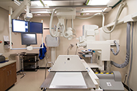
X-ray
What are X-Rays?
 X-ray imaging (radiography) is still the most commonly used technique in radiology. To make a radiograph, a part of the body is exposed to a very small quantity of X-rays. The X-rays pass through the tissues, striking a film to create an image. X-rays are safe when properly used by radiologists and technologists specially trained to minimize exposure. No radiation remains after the radiograph is obtained.
X-rays are used to image every part of the body and are used most commonly to look for fractures. They are also commonly used to examine the chest, abdomen, and superficial soft tissues. X-rays can identify many different conditions within the body, and they are often a fast and easy method for your doctor to make a diagnosis.
What Should I Expect?
X-rays are fast, easy, and painless. The part of your body to be examined will be properly positioned, and several different views of that part may be obtained. The technologist will instruct you to hold still and in some cases hold your breath while the X-ray is being taken to eliminate blurring. X-ray exams generally take around 20 minutes, after which you will be able to return to normal activities.
How Should I Prepare?
Preparation is not required for an x-ray. You may be asked to change into a hospital gown to eliminate the chance of artifacts from your clothing. You will also be asked to remove any jewelry, eyeglasses, or any other metal objects. Women should always inform their technologist if there is any possibility of pregnancy.
How Do I Get the Results?
After your study is over, the images will be evaluated by one of our board-certified radiologists. A final report will be sent to your doctor, who can then discuss the results with you in detail.
Appointments
Appointments for the Providence Imaging Center and the Providence Medical Center Radiology department can be made at 913-667-5600
For more information please visit www.Radiologyinfo.org
Featured Services
Emergency Services

Heart and Vascular
Maternity Care
Neurosciences
Orthopedics


