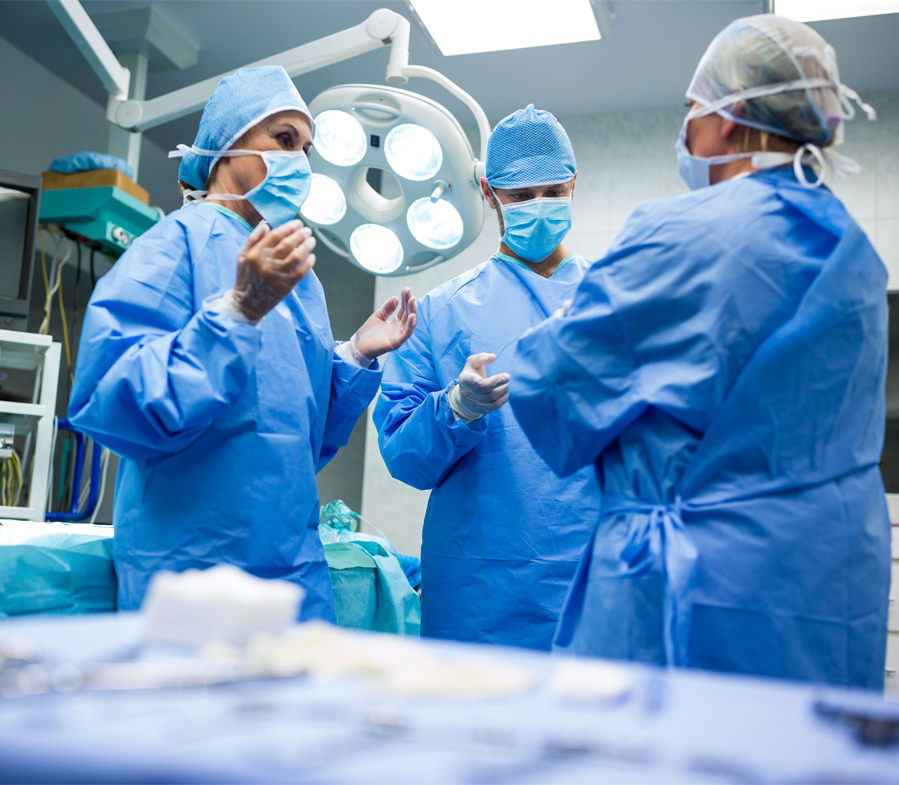
Vascular Surgery
More than eight million Americans suffer from peripheral vascular disease (PVD).
PVD refers to diseases of the blood vessels outside of the heart, such as those that carry blood to the brain, legs, arms, stomach or kidneys.
It occurs when arteries narrow or clog. People often refer to this as hardening of the arteries.
Studies estimate that only half of those with PVD are seeing a doctor for treatment. Find out how you can obtain a free PAD Screening.
Providence Medical Center has one of the area’s only surgical teams devoted solely to diagnosing and treating vascular diseases.
For further information, contact us at 913-600-4908
Risk Factors for PVD
PVD is more common among those over 50 and affects from 12 to 20 percent of those age 65 and older. The population in Wyandotte and Leavenworth counties has a high incidence of the conditions that make people susceptible to PVD, such as diabetes and high blood pressure. Here’s how to determine if you are at risk for PVD:
- Do you smoke?
- Do you have cardiovascular (heart) problems such as high blood pressure, heart attack or stroke?
- Do you have high cholesterol?
- Do you have a family history of diabetes or cardiovascular problems (immediate family such as parent, sister, brother)?
- Do you have diabetes?
- Are you more than 25 pounds overweight?
- Do you have an inactive lifestyle?
If you answered “Yes” to any, you are at risk. More than two signals high risk.
For more information, talk with your primary care physician or call the Providence Vascular Surgery team at 913-262-9201.
Symptoms of PVD
PVD often goes undiagnosed because many don’t have symptoms in the early stages or they mistakenly think the symptoms are a normal part of aging. About half of those with PVD will have some symptoms, such as:
- Aching, cramping or pain in your calves or thighs when you walk or exercise. The pain typically goes away when the muscles are given a rest.
- Pain in your toes or feet at night
- Sores on your leg or foot that are slow to heal.
- Extreme cases can lead to gangrene, a serious condition that may require amputation of a leg, foot or toes.
Treating PVD
The Providence Vascular Surgery team provides a full range of care from prevention to surgery. Frequently, lifestyle changes, such as weight loss, dietary changes, smoking cessation and more exercise, will prevent or stop PVD from progressing. Medical therapy can also slow or stop the disease process. This can involve medications to control blood pressure, manage blood lipid levels and specific medications for vascular disease, as well as daily aspirin.
Cases that are more serious may require intervention. Medical advances often allow our surgeons to treat PVD with minimally invasive procedures that require only tiny incisions and allow you to recover in hours as opposed to weeks. They use small catheters (tubes) or other devices, which they guide by X-ray or ultrasound imaging to the affected areas. These procedures are usually less traumatic for patients than conventional surgery. They involve minimal pain, shorter (or no) hospital stays, allow faster recovery and often cost less than invasive surgery.
If necessary, our team can perform traditional open surgery.
Here are some of the techniques our Vascular Surgery team uses to treat PVD:
Angioplasty – Surgeons place a tiny balloon in the blood vessel at the site of the blockage. Then, they inflate it to open the blood vessel.
[Visit WWW.VASCULARSURGERYASSOC.NET to see a balloon angioplasty image]
Stents – Surgeons insert a tiny metal cylinder, or stent, in the clogged vessel to act like scaffolding and hold it open.
[Visit WWW.VASCULARSURGERYASSOC.NET to see an image of a Stent]
Atherectomy – Surgeons use a rotating shaver at the end of a catheter to shave off plaque.
Bypass grafts – Surgeons take a vein graft from another part of the body or one made from artificial material to create a detour around a blocked artery. This technique can restore circulation in arteries to the brain, kidneys or legs.
Thrombolytic therapy – When symptoms of PVD develop suddenly, as a result of a blood clot, a catheter is used to deliver clot-busting drugs directly to the site of the clot that is causing a blockage. Doctors frequently combine these drugs with another treatment, such as angioplasty.
Thrombectomy – This is another way to treat blood clots. Surgeons insert a balloon catheter into the affected artery beyond the clot. They inflate the balloon and pull it back, bringing the clot with it.
Open surgery – This may be required to remove blockages from arteries or to bypass the clogged area.
Combined approach – The Vascular Surgery team sometimes combines techniques, using minimally invasive procedures along with traditional surgery to treat multi-level vascular disease simultaneously.
Procedures We Preform
Carotid Endarterectomy
Carotid arteries carry blood to the brain. When they narrow or clog, your brain can’t get enough oxygen. Plaque buildup also makes the wall of the artery rough. Plaque can then break loose and flow into the brain causing a stroke or TIA (a transient ischemic attack or “mini stroke”). For this procedure, surgeons make a small incision in your neck and open the carotid artery. Then they remove the plaque to make the artery smooth. This procedure is currently the treatment associated with the lowest risk of complications for removing blockages. It typically requires a one-day hospital stay. Most patients are able to resume normal activities within a week. Surgeons also use a similar type of procedure in other areas of the body. [Visit WWW.VASCULARSURGERYASSOC.NET to view an image of a carotid artery diagram.]
Carotid Stenting
Some patients aren’t good candidates for open surgery. Those with severe lung or heart disease or who have had prior carotid surgery or neck irradiation may be treated with stents. Surgeons insert a stent through a remote site, such as the femoral artery, for access. They then thread it through a catheter to the carotid artery to dilate the blockage.
Endovascular Aneurysm Repair
An abdominal aortic aneurysm (AAA) is a weak area in the main blood vessel that carries blood from the heart to the rest of the body. It causes the aorta to bulge like a balloon and can rupture if it gets too big. If it does, your chances of survival are low; only 25 percent will survive a ruptured aneurysm. In the past 30 years, the occurrence of these has increased threefold. AAAs affect as many as eight percent of people over the age of 65. AAA repair used to require open surgery and more than a week in the hospital. For an endovascular repair, surgeons insert a catheter through small incisions over the groin arteries. They use this to thread a reinforced fabric tube to seal off the aneurysm. Most patients go home after a day or two in the hospital. [Visit WWW.VASCULARSURGERYASSOC.NET to view image.]
Surgical Aneurysm Repair
Surgeons make an incision in the abdomen and clamp the artery above and below the aneurysm to stop the flow of blood into the area. Then they replace the aneurysm segment with a synthetic graft. This procedure usually requires a five-to-seven day hospital stay. Full recovery usually takes several weeks after the patient goes home.
Varicose Vein Ablation
Your veins carry blood from the capillaries to the heart. In your legs, the blood has to flow upward against gravity. Consequently, the veins have one-way valves to prevent back flow. Over time, these valves can fail to close tightly, allowing blood to pool. This causes the bulging and twisting characteristics of varicose veins. In mild conditions, compression stockings can help, but, if the problem grows more severe, the abnormal vein must be eliminated. Historically, this has been done with a surgical procedure called “vein stripping.” Now, it can be done with venous ablation. Your surgeon makes a tiny incision at the knee, inserts a small tube in the vein, then passes a laser or concentrated beam of light to the problem area. There it delivers localized heat to the vein wall. In response, the vein closes down and becomes permanently blocked. It takes about 45 minutes and requires just local anesthesia. Patients usually see results soon after the surgery. The procedure has proven to be 97 percent effective in eliminating varicose veins.
Spider Vein Treatment (Sclerotherapy)
Spider veins are the thin strands of red, blue or purple veins close to the skins surface. They typically form on your legs or thighs. Unlike varicose veins, these don’t bulge. Heredity, pregnancy, weight gain or prolonged standing can cause them. While spider veins don’t cause any health problems, many find them unsightly. Surgeons can diminish them by injecting a caustic substance directly into the affected area. This shuts off the tiny veins, and the body reabsorbs the tissue. Sclerotherapy doesn’t require any anesthesia and patients can quickly resume normal activities, although it takes about a month to achieve the final results.
Vascular Access for Kidney Patients
Patients who must rely on dialysis to filter blood wastes need special access to their veins, such as a fistula, graft or catheter. This procedure is done on an outpatient basis using conscious sedation and local anesthesia.
For more information about PVD click Society for Vascular Surgery
Featured Services
Emergency Services

Heart and Vascular
Maternity Care
Neurosciences
Orthopedics


